

What does the retina do?
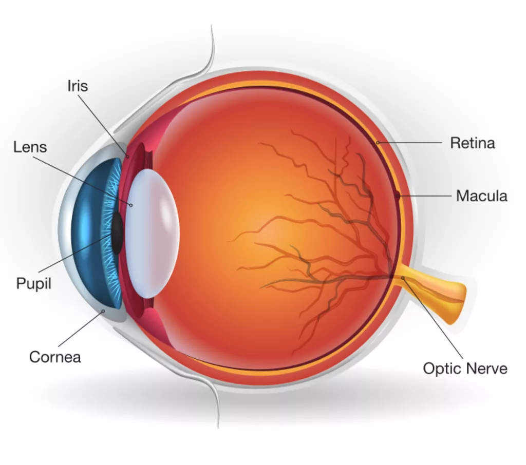
The retina is the light-sensitive nerve tissue lining the back of the eye. Like film in a video camera, the retina continually ‘take pictures’, then sends signals through the optic nerve to the brain, producing vision. The central part of the retina, which generates fine detailed vision, is known as the macula. The retina is nourished by oxygen-rich blood that is brought to it be arteries and then drained away again by veins.
What is a retinal vein occlusion?
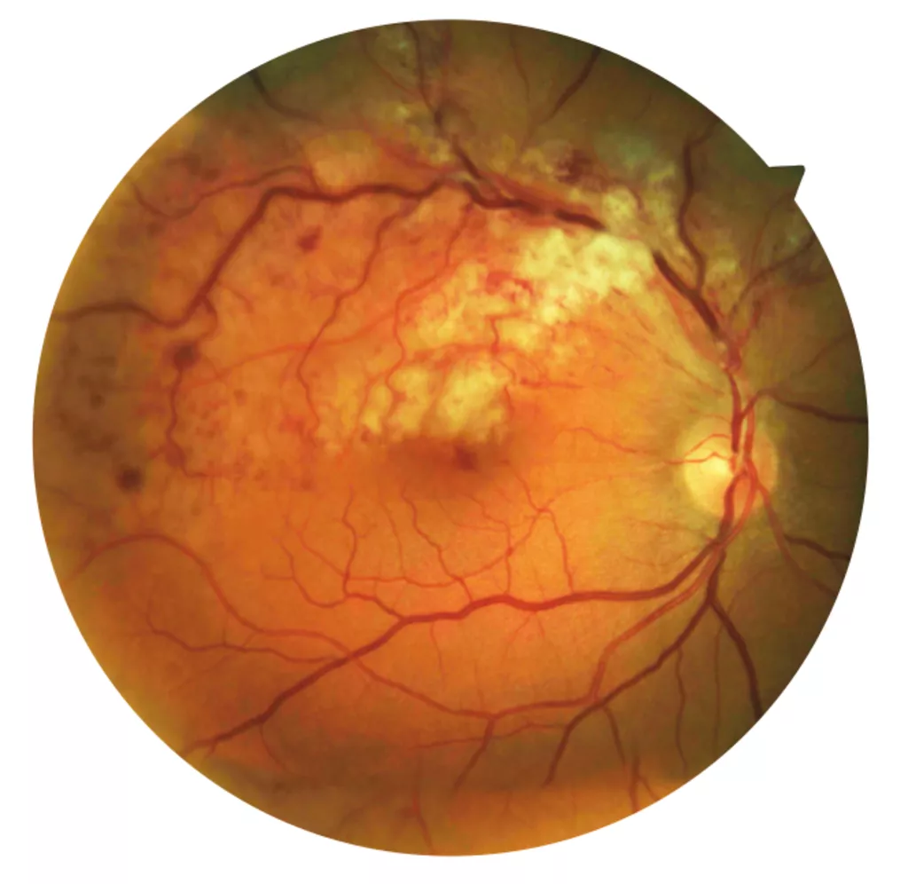
When a retinal vein becomes blocked, part of the retinal blood flow slows or stops. Blood backs up, leading to bleeding and swelling of the retina, hampering its nourishment. Suddenly, and usually without warning, a patch of retina loses some of its ‘picture-taking’ function.
What are the main symptoms of a retinal vein occlusion?
The retinal swelling caused by a blocked vein can lead to a sudden loss of vision, or distortion of the central vision, which are often the first signs that many people notice. The symptoms vary from none at all to almost total loss of vision, depending on the type and severity of the vein occlusion.
When the Central Retinal Vein becomes blocked, the whole retina is affected, which can cause severe symptoms. When a Branch Vein becomes blocked, symptoms can be minor if the periphery of the retina is affected, but more severe if the central area of the macular is involved.
What causes a vein occlusion?
Vein occlusions become more common with age and as blood vessels harden. Risk factors that ‘age’ the veins faster can include smoking, diabetes, high blood pressure and high cholesterol. Glaucoma and inflammation inside the eye can also increase the risk of a blockage.
What examinations are required?
Your eye surgeon will check your vision and examine the back of the eye. The pupil will be dilated with drops, so you will not be able to drive for a few hours. Photos may be taken with a camera and a special OCT (Optical Coherence Tomography) scan will be used to map the back of the eye and demonstrate any swelling in the retina. Sometimes further tests, such as Fluorescein angiography may be required, where dye is injected into an arm vein and is then photographed as it travels through to the back on the eye.
What treatment can help with retinal vein occlusion?
Depending
on the severity and position of the occlusion, often no treatment is
required. However sometimes we can speed the recovery by using
medications that target the leaking vessels. These medications can
include injections into the eye, which may need to be repeated. Laser
treatment may also be required to close down leaking vessels in
affected parts of the retina.
With time your own healing process
may open up the blocked vessel. The resulting vision will depend on
the amount of damage to the retina. The treatment is tailored to the
individuals needs to give the best chance of regaining long term
vision.